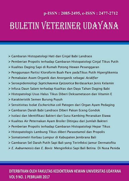
PROFILE OF WHITE BLOOD CELLS IN BALI CATTLE NATURALLY INFECTED BY DERMATOPHYTOSIS
Abstract
Dermatophytosis is kind of disease which caused by dermatophyta fungus. White blood cell (leukocyte) will responds every strange things which entering the body as a defend cell. In Indonesia, only few information of the white blood cell (leukocyte) in the Dermatophytosis cases towards balinese cattle can be found. The purpose of the research in finding the comparasion between lukocyte of normal balinese cattle which is not infected and balinese cattle which infected by dematophyta fungus. The research are using 12 samples of blood, is abaout 6 blood’s sampel from normal balinese cattle and 6 blood’s sample of balinese cattle which is infected by dermatophyta fungus. The first attempt is checking the skin scratching and the hair with 10% of KOH liquid. Sabouraud’s Dextrose Agar (SDA) is used for isolate and identified dermatophyta fungus. Calculation and checking the total of leukocyte are using hemasitometer, while Giemsa liquid are using for differential leukocyte. T-Test shows the real differences between them which in to balinese cattle which infected by dermatophytosis. The analyzes result bye the statistic data using Mann-Whitney Test is showing there is real difference to balinese cattle’s monocyte which infected by dermatofitosis. There is a normal difference between balinese cattle which is normal and infected. Balinese cattle which infected dermatophyta fungus have a total of leukocyte and monocyte are higher than normal balinese cattle.
Downloads
References
Ainsworth GC. 1986. Introduction to the History Of Medical and Veterinary Mycology. Cambridge University Press. Cambridge, London, New York, New Rochelle, Melbourne, Sydney. 228p.
Bandini Y. 2004. Sapi Bali. Penebar Suadaya. Jakarta.
Besung INK. 2009. Pegagan (Centella asiatica) sebagai alternatif peneguhan penyakit infeksi pada ternak. Bul Vet Udayana 1(2): 99-105.
Blanco JL, Gracia ME. 2008. Immune response to fungal infections. Vet Immunol Immunopathol 125: 47-70.
Charles AJ. 2002. Immunobiology, Chapter 11: Failures of Host Defense Mechanisms. (Diakses pada 15 Maret 2010).
Daniela C, Rapuntean Gh, Nicodim FIT, George N, Ioan M, Iulia P, dan Romana-Maria O. 2009. Evaluation of the cellular non-specific defense effectors in cattle ringworm vaccination. Bull Univ Agric Sci Vet Med Cluj-Napoca, Vet Med 66 (1): 266-272.
Djaenudin G, Rachmawati S. 2010. Kapang dermatofita Trichopyton verrucosum penyebab penyakit ringworm pada sapi. Balai Besar Veteriner. Bogor. pp: 1-11.
Dharmawan NS. 2002. Pengantar Patologi Klinik Veteriner Hematologi Klinik. Universitas Udayana Kampus Bukit Jimbaran. Bali.
Guyton, Hall. 1997. Buku Ajar Fisiologi Kedokteran. Ed ke-9. Penerjemah Irawati Setiawan. Jakarta:EGC.
Hughes NC, Wickramasinghe SN. 1995. Catatan Kuliah Hematologi Penerbit Buku Kedokteran EGC. Jakarta.
Jain LC dan Musc AH. 1986. Schalm’s Veterinary Hematology. Lea & Fibiger. Philadelphia. pp: 450-500.
Kartono A, Rosidah, Arif A. 2010. Solusi numerik model dinamik perlakuan immunotherapy pada infeksi HIV-1. Berkala Fisika 13(1): 1-10.
Koga TH, Duan K, Urabe, Furue M. 2001. Immunohoistochemical detection of interferon-gamma producing cells in dermatophytosis. Eur J Dermatol 11(2): 105-107.
Kotik T, Corne M. 2006. Clinical and histopathhological evaluation of terbinafine treatments in cats experimentally infected with Microsporum canis. Acta Vet 75: 541-547.
Leijht PCJ, Furh V, Zweet TLV. 1986. In Vitro Determination of Phgocyte and Intracellular Killing by Polymorphonuclear and Intracellular Phagocyte. In Weir DM. Ed Cellular Immunology, Blackwell Scientific Publication. London. pp: 1-46.
Meyer DJ, Harvey JH. 2004. Veterinary Laboratory medicine: Interpretation and diagnosi. 3rd Ed. Saunders.
Mignon BR, Leclipteux T, Focant CH, Nikkels AJ, Pierard GE, Losson BJ. 1999. Humoral and cellular immune respone to a crude exoantigen and purified keratinase of microsporum canis in experimentally infected guinea pigs. Med Mycol 37: 123-129.
Mignon BR, Leclipteux T, Focant CH, Nikkels AJ, Pierard GE, Losson BJ. 2008. Humoral and cellular immune respone to a crude exoantigen and purified keratinase of microsporum canis in experimentally infected guinea pigs. Med Mycol 37: 200-205.
Matrsuda Y, Namikawa T, Kondo, Martojo H. 1980. A study on karyotypes of the bali cattle. The origin and phylogeny of Indonesia native lives stock. 29-33.
Pradana IMYW, Sampurna IP, Suatha IK. 2014. Pertumbuhan dimensi tinggi tubuh pedet sapi bali. Bul Vet Udayana 6(1): 81-85.
Siswanto. 2011. Gambarab sel darah merah sapi bali (Studi Rumah Potong). Bul Vet Udayana 3(2): 99-105.
Sohnle PG. 1993. Dermatophytosis In: Murphy, Friedman JW, Bendinello H. Editor Fungal Infection and Immune Respone. New York: Plenum. New York. pp: 27-47.
Sparkes AHG, Jones TJ, Shaw SE, Wright AI, Stokes CR. 1993. Epidemiologi dan diagnostic feature of canine and feline dermatophytosis in united kingdom from 1956 to 1991. Vet Rec pp: 135-142.
Utama IH, Kendra AAS, Widyastuti SK, Virginia P, Sene SM, Kusuma WD, Arisdani BY. 2013. Hitungan differensial dan kelainan sel darah sapi bali. J Vet 14(4): 462-466.
Vermount S, Tabart J, Baldo A, Mathy, Losson B, Mignan B. 2008. Pathogenesis of dermatophytosis. Mycopathologia 54: 299-308.
Wisesa AANGD, Pemayun TGO, Mahardika IGNK. 2012. Analisis sekuens D-Loop DNA mitokondria sapi bali dan banteng dibandingkan dengan bangsa sapi lain di dunia. Indon Med Vet 1(2): 281-192.
Zukesti E. 2003. Peranan leukosit sebagai anti inflamasi alergik dalam tubuh. USU Library (Diakses pada 20 juni 2015).


