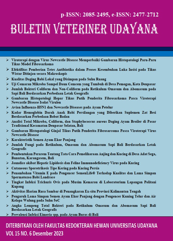
TRICHURIS OVIS INFECTION RATES IN THE DRY SEASON IN THE KUPANG POLITANI FIELD LABORATORY
Abstract
The goats being raised semi intensively on the field laboratory of State Agricultural Polytechnic of Kupang were kacang goats (Capra hircus). Objective: to measure Trichuris ovis infectious levels based on age during dry season. Nine heads of goats were grouped into two of four for 6 to 7 and five for 18 to 24 month old. Two methods to identify morphological worm eggs are sedimentation and floating; while infectious intensity was using McMaster method. Faecal samples for the two groups were collected twice a week. Descriptive analysis was applied to determine gastrointestinal endoparasites and endoparasite infectious intensity, so that of ambient temperature of pasture. The results showed that average infectious intensity of Trichuris ovis worm eggs for 6 to 7 month old goats was 100 TTG while that for 18 to 24 month old was 200 TTG. It can be concluded that the intensity of Trichuris ovis infection in goats aged 18 to 24 months is higher than that aged 6 to 7 months and is included in the light infection category. This infection category is influenced by season and food source. The difference in the age of the goats is one of the factors causing the difference in the intensity of Trichuris ovis infection between the two groups. The author suggests that in further Res. it would be better to use a larger number of samples with a longer Res. time so that the results are more accurate.
Downloads
References
Amaral AC, Freitas JDC, de Carvalho RD, Noronha AMDCG, Ribeiro JMDS, Santos ID. 2022. The prevalence of Strongylida/strongyles in small ruminants in Manatuto Municipality in central region of Timor-Leste. Livestock Anim. Res.. 20(2): 110-117.
Anggraini M, Primarizky H, Mufasirin, Suwanti LT, Hastutiek P, Koesdarto S. 2019. Prevalensi penyakit protozoa darah pada sapi dan kerbau di Kecamatan Moyo Hilir Kabupaten Sumbawa Nusa Tenggara Barat. J. Parasite Sci. 3(1): 9-14.
Beriso G, Tesfaye Z, Fesseha H, Asefa I, Tamirat T. 2023. Study on gastrointestinal nematode parasite infections of donkey in and around shone town, Hadiya zone, Southern Ethiopia. Heliyon. 9(6): e17213.
Bowman DD, Georgi JR. 2009. Georgi’s parasitology for veterinarians. Elsevier Health Sciences: United Kingdom.
Bulbul KH, Akand AH, Hussain J, Parbin S, Hasin D. 2020. A brief understanding of Trichuris ovis in ruminants. Int. J. of Vet. Sci. Anim. Husb. 5(3): 72-74.
Crotti M. 2013. Digenetic Trematodes: an existence as parasites. Brief general overview. Microbiol. Med. 28(2): 97-101.
Hussein HA, Abdi SM, Ahad AA, Mohamed A. 2023. Gastrointestinal nematodiasis of goats in Somali pastoral areas, Ethiopia. Parasite Epidemiol. Control. 23: 1-7.
Jabbar A, Gauci C, Lightowlers MW. 2016. Diagnosis of human taeniasis. Faculty of Vet. and Agricultural Sciences, The University of Melbourne, Werribee, Vic. 3030, Australia. Page. 43-45.
MAFF. 1986. Fisheries and food, reference book, manual of Vet. parasitological laboratory techniques, Vol.418, Ministry of Agriculture, HMSO, London, 5pp
Muhajirin M, Despal D, Khalil K. 2017. Pemenuhan kebutuhan nutrien sapi potong bibit yang digembalakan di Padang Mengatas. Bul. Makanan Ternak. 104(1): 9-20.
Mpofu TJ, Nephawe KA, Mtileni B. 2022. Prevalence and resistance to gastrointestinal parasites in goats: a review. Vet. World. 15: 2442-2452.
Nurcahyo RW, Ekawasti F, Haryuningtyas D, Wardhana AH, Firdausy LW, Priyowidodo D, Prastowo J. 2021. Occurrence of gastrointestinal parasites in cattle in Indonesia. IOP Publishing. doi:10.1088/1755-1315/686/1/012063.
Orden EA, Rosario NAD, Orden MEM, Fujihara T. 2017. Nutritive value and anthelmintic properties of selected leguminous shrubs and trees for goats. Int. J. Sci. Technol. 2(2): 28-37.
Paul BT, Jesse FFA, Chung ELT, Che’Amat A, Lila MAM. 2020. Risk factors and severity of gastrointestinal parasites in Selected Small Ruminants from Malaysia. Vet. Sci. 7(208): 1-14.
Shakya P, Jayraw AK, Jamra N, Agrawal V, Jatav GP. 2017. Incidence of gastrointestinal nematodes in goats in and around Mhow, Madhya Pradesh. J. Parasit Dis. 41(4): 963-967.
Silvestre R-C, Camalig FeM, Juliana Q. 2013. Prevalence of ectoparasites and endoparasites of carabao in Region I. E – Int. Sci. Res. J. 5(3).
Tiele D, Sebro E, Meskel DH, Mathewos M. 2023. Epidemiology of gastrointestinal parasites of cattle in and Around Hosanna Town, Southern Ethiopia. Vet. Med. 14: 1 – 9.
Urquhart GM, Armour J, Ducan JL, Dunn AM, Jennings FW. 1987. Vet. parasitology. Longman Group UK Limited. Published by Churchill Livingstone Inc., New York, USA, 92-93.
Wirawan IGKO, Nurcahyo W, Prastowo J, Kurniasih K. 2017. Daya larvasida ekstrak daun muda kedondong hutan terhadap Haemonchus contortus Secara In-vitro. J. Vet. 18(2): 283-288.
Wirawan IGKO, Jaya IK, Randu MDS. 2019. Keragaman dan intensitas infeksi endoparasit gastrointestinal pada sapi bali dengan sistem ekstensif di Kabupaten Kupang. J. Sain Vet. (37)2: 151-159.
Wirawan IGKO, Suryawati S, Koni TNI, Wea R. 2022. Daya vermisidal ekstrak air dua jenis etnofarmakologi terhadap Haemonchus contortus pada kambing kacang (Capra hircus) secara In-vitro. J. Sain Vet. 40(2): 147-154.
Zajac AM, Conboy GA. 2012. Vet. clinical parasitology. 8th Edition. American Association of Vet. Parasitologists. ©Iowa State University Press.
Zalizar L. 2017. Helminthiasis saluran cerna pada sapi perah. J. Ilmu-Ilmu Pet. 27(2): 116-122.


