Brain Tumor Segmentation Based on Magnetic Resonance Imaging Images Using the U-NET Method
Abstract
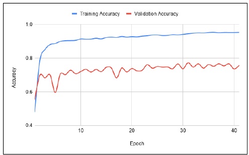
Brain tumor is a deadly disease where 3.7% per 100,000 patients have malignant tumors. To analyze brain tumors can be done through magnetic resonance imaging (MRI) image segmentation. Automatic image analysis process is needed to save time and improve accuracy of doctor diagnoses. Automatic segmentation can be done with deep learning. U-NET is one of the methods used to segment medical images because it works at pixel level. By applying the ReLU and Adam Optimizer activation function, this method can solve the problem of segmenting brain tumors. Dataset for the training and validation process using BRATS 2017. Several hyperparameters are applied to this method: learning rate (lr) = 0.0001, batch size (bz) = 5, epoch = 80 and beta (b_1) = 0.9. From a series of processes carried out, accuracy of the U-NET method is calculated by Dice Coefficient formula and results in following accuracy values, during training of 90.22% (Full Tumor), 78.09% (Core Tumor) dan 80.20% (Enhancing Tumor).
Downloads
References
Brain Tumor Epidemiology Consortium (BTEC),” NIH Public
Access, vol. 113, no. 1, pp. 1953–1968, 2010.
[2] R. Riley, J. Murphy, and T. Higgins, “MRI imaging in pediatric
appendicitis,” J. Pediatr. Surg. Case Reports, vol. 31, no. January,
pp. 88–89, 2018.
[3] I. Bagus, L. Mahadya, R. S. Hartati, and Y. Divayana, “Diagnosa
Tumor Otak Berdasarkan Citra MRI ( Magnetic Resonance
Imaging ),” Maj. Ilm. Teknol. Elektro, vol. 18, no. 2, pp. 149–154,
2019.
[4] Tech Wire Asia, “Medical diagnosis AI in Beijing beats real
doctors,” Tech Wire Asia, 2018. [Online]. Available:
https://techwireasia.com/2018/07/medical-diagnosis-ai-in-beijingbeats-
real-doctors/.
[5] P. P. Winangun, I. M. O. Widyantara, and R. S. Hartati, “Learning
Machine dengan Kernel Linear untuk Mengklasifikasi Kelainan
Paru-Paru,” Maj. Ilm. Teknol. Elektro, vol. 19, no. 1, pp. 83–88,
[6] A. Pinto, H. Correia, J. Oliveira, D. M. L. D. Rasteiro, and C. A.
Silva, “Brain Tumour Segmentation based on Extremely
Randomized Forest with High-Level Features,” pp. 3037–3040,
2015.
[7] N. Subbanna, D. Precup, and T. Arbel, “Iterative multilevel MRF
leveraging context and voxel information for brain tumour
segmentation in MRI,” Proc. IEEE Comput. Soc. Conf. Comput. Vis.
Pattern Recognit., pp. 400–405, 2014.
[8] M. Soltaninejad et al., “Automated brain tumour detection and
segmentation using superpixel-based extremely randomized trees in
FLAIR MRI,” Int. J. Comput. Assist. Radiol. Surg., vol. 12, no. 2,
pp. 183–203, 2017.
[9] W. Wu, A. Y. C. Chen, L. Zhao, and J. J. Corso, “Brain tumor
detection and segmentation in a CRF (conditional random fields)
framework with pixel-pairwise affinity and superpixel-level
features,” Int. J. Comput. Assist. Radiol. Surg., vol. 9, no. 2, pp.
241–253, 2014.
[10] S. Shen, W. A. Sandham, M. H. Granat, M. F. Dempsey, and J.
Patterson, “A New Approach to Brain Tumour Diagnosis Using
Fuzzy Logic Based Genetic Programming,” in Proceedings of the
25th Annual International Conference of the IEEE EMBS, 2004, vol.
1, pp. 870–873.
[11] A. Das and M. Bhattacharya, “A study on prognosis of brain tumors
using fuzzy logic and genetic algorithm based techniques,” in
Proceedings - 2009 International Joint Conference on
Bioinformatics, Systems Biology and Intelligent Computing, IJCBS
2009, 2009, no. 1, pp. 348–351.
[12] L. Szilagyi, L. Lefkovits, and B. Benyo, “Automatic Brain Tumor
Segmentation in multispectral MRI volumes using a fuzzy c-means
cascade algorithm,” 2015 12th Int. Conf. Fuzzy Syst. Knowl. Discov.
FSKD 2015, pp. 285–291, 2016.
[13] J. Belghese and S. Agustin, “Brain Tumor Segmentation using
Pattern Neural Networks with MRI Images,” IJSTE -International J.
Sci. Technol. Eng., vol. 3, no. 09, pp. 641–644, 2017.
[14] S. Singh, “Classification of Human Brain Tumors from MRI Using
K-NN Algorithm,” Int. J. Adv. Sci. Eng. Technol., vol. 3, no. 1, pp.
34–37, 2015.
[15] R. M. Azawi and I. T. Ibrahim, “A Hybrid Approach for
Classification of MRI Brain Tumors Using Genetic Algorithm , KNearest
Neighbor and Probabilistic Neural Network,” Int. J.
Comput. Sci. Inf. Secur., vol. 16, no. 5, pp. 74–85, 2018.
[16] A. Arora, P. Roy, S. Venktesan, and R. Babu, “k-NN Based
Classification of Brain MRI Images using DWT and PCA to Detect
Different Types of Brain Tumour,” Int. J. Med. Res. Heal. Sci., vol.
6, no. 9, pp. 15–20, 2017.
[17] W. Mengqiao, Y. Jie, C. Yilei, and W. Hao, “The multimodal brain
tumor image segmentation based on convolutional neural networks,”
2017 2nd IEEE Int. Conf. Comput. Intell. Appl., pp. 336–339, 2017.
[18] S. Hussain, S. M. Anwar, and M. Majid, “Brain Tumor
Segmentation using Cascaded Deep Convolutional Neural Network,”
39th Annu. Int. Conf. IEEE Eng. Med. Biol. Soc., pp. 1998–2001,
2017.
[19] A. Pinto, V. Alves, and C. A. Silva, “Brain Tumor Segmentation
using Convolutional Neural Networks in MRI Images,” IEEE Trans.
Med. Imaging, vol. 35, no. 5, pp. 1240–1251, 2016.
[20] J. W. Ha et al., “Predicting high-risk prognosis from diagnostic
histories of adult disease patients via deep recurrent neural
networks,” 2017 IEEE Int. Conf. Big Data Smart Comput. BigComp
2017, pp. 394–399, 2017.
[21] O. Ronneberger, P. Fischer, and T. Brox, “U-net: Convolutional
networks for biomedical image segmentation,” Lect. Notes Comput.
Sci. (including Subser. Lect. Notes Artif. Intell. Lect. Notes
Bioinformatics), vol. 9351, pp. 234–241, 2015.
[22] L. Dirven, N. K. Aaronson, J. J. Heimans, and M. J. B. Taphoorn,
“Health-related quality of life in high-grade glioma patients,” Chin.
J. Cancer, vol. 33, no. 1, pp. 40–45, 2014.
[23] W. Rawat, “Deep Convolutional Neural Networks for Image
Classification : A Comprehensive Review,” vol. 2449, pp. 2352–
2449, 2017.
[24] G. Ö. Yiğit and B. M. Özyildirim, “Comparison of convolutional
neural network models for food image classification,” vol. 1839,
2018.
[25] N. Sharma, V. Jain, and A. Mishra, “An Analysis of Convolutional
Neural Networks for Image Classification,” Procedia Comput. Sci.,
vol. 132, no. Iccids, pp. 377–384, 2018.
[26] N. M. Balasooriya and R. D. Nawarathna, “A sophisticated
convolutional neural network model for brain tumor classification,”
2017 IEEE Int. Conf. Ind. Inf. Syst., pp. 1–5, 2017.

This work is licensed under a Creative Commons Attribution-NonCommercial-NoDerivatives 4.0 International License.

This work is licensed under a Creative Commons Attribution 4.0 International License